In the dynamic field of reproductive endocrinology and infertility (REI), even experienced practitioners from different specialties may struggle to navigate embryo reports. While most of us remember the fundamentals of mitosis and meiosis from our medical training, the specific clinical details of embryo morphology and grading are frequently not covered until fellowship.
Grasping these metrics is crucial for making informed choices. Whether you are an OB/GYN setting patient expectations or a clinician undergoing IVF personally, here is a practical, evidence-driven guide to embryo grading.
The initial phases: Days 0 to 3
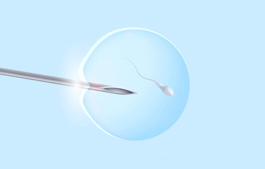
When eggs are collected during a transvaginal ultrasound-guided retrieval (Day 0), the lab works with a predetermined number of oocytes. These eggs are either subjected to sperm (standard insemination) or injected with a single sperm (intracytoplasmic sperm injection, or ICSI). A fertilization assessment occurs roughly 18 hours later.
By Day 1, the aim is to observe an embryo featuring two pronuclei (one from the mother and one from the father), termed a 2PN embryo. Differences may include immature eggs or embryos with 0PN, 1PN, or 3PN or more. Since each pronucleus has a haploid collection of chromosomes, deviations from 2PN raise concerns about abnormal ploidy. Nevertheless, due to potential mis-timings in fertilization, embryos are typically kept in culture to evaluate later development.
By Day 3, embryos should have experienced rapid division, with a goal of reaching the eight-cell phase. Day 3 grading incorporates a number and a letter. The number signifies cell quantity (e.g., 6, 8, or 10 cells). Even-numbered embryos generally indicate more favorable outcomes, although odd-numbered embryos can successfully lead to live births. The letter denotes morphology: “A” or “B” grades suggest minimal fragmentation and cohesive blastomeres, while “C” or “D” grades indicate inferior morphology.
Between Days 3 and 5, embryos move through the morula phase (a compact cluster of cells that is hard to evaluate). Consequently, most labs do not officially grade embryos on Day 4.
Comprehending blastocyst grading
By Day 5 or 6, embryos ideally achieve the blastocyst stage, identified by a fluid-filled cavity (the blastocele), an inner cell mass (ICM), and the trophectoderm (TE). Some labs may prolong culture to Day 7, though implantation rates typically decrease due to delayed maturation.
Blastocyst grading consists of one number and two letters (e.g., 4AA). The number indicates the extent of expansion and hatching from the zona pellucida, rather than cell count. Grades 1 and 2 denote early blastocysts with less defined ICM and TE layers. Grade 3 is a fully developed blastocyst with distinct layers. Grade 4 represents an expanded blastocyst with thinning zona pellucida. Grade 5 describes an actively hatching embryo, and Grade 6 a completely hatched embryo. Notably, a Grade 6 embryo isn’t necessarily “better” than a Grade 4; what’s significant is viability, expansion, and developmental progress. When advising patients, I often characterize this stage as the embryo actively “seeking a home.”
The first letter evaluates the ICM (the group of cells destined to form the fetus), while the second letter assesses the TE, which creates the placenta. As with most grading criteria, A is better than B, which is better than C. The TE grade holds clinical relevance, as these cells facilitate implantation and initial nutrient exchange.
Debate continues regarding the significance of ICM compared to TE grade and whether Day 5 embryos are preferable to Day 6 embryos. Individual labs possess their own outcome statistics, but to this day, no conclusive answers have resolved these inquiries.
Beyond morphology: Genetic testing
While morphology assists in prioritizing embryos for transfer, it doesn’t provide the complete picture. A high-quality embryo can be aneuploid, and a lower-graded embryo can lead to a healthy, euploid live birth.
Numerous laboratories now provide Preimplantation Genetic Testing for Aneuploidy (PGT-A), which consists of biopsying several trophectoderm cells at the blastocyst stage. At this juncture, embryos comprise approximately 100–200 cells. Although PGT-A is broadly adopted, its advantages remain complex. Evidence indicates that in patients under 38 without a history of recurrent pregnancy loss, PGT-A may not enhance outcomes and could even be harmful due to increased embryo manipulation. Conversely, for patients over 38 or those with recurrent aneuploid losses, PGT-A can boost pregnancy rates per transfer, minimize miscarriage risks, reduce twin rates with single embryo transfers, and shorten the time to live birth.
Nevertheless, growing recognition of mosaicism and variability among testing methods has moderated initial excitement. Interpreting mosaic,